Last update images today Decoding Your Feet: An Anatomy Image Guide
Decoding Your Feet: An Anatomy Image Guide
This week, let's delve into the fascinating world of foot anatomy, exploring the intricate network of bones, muscles, ligaments, and nerves that allow us to stand, walk, run, and dance! Understanding your foot anatomy image can empower you to better care for your feet, prevent injuries, and even improve your athletic performance.
Understanding the Bones: Foot Anatomy Image in Detail
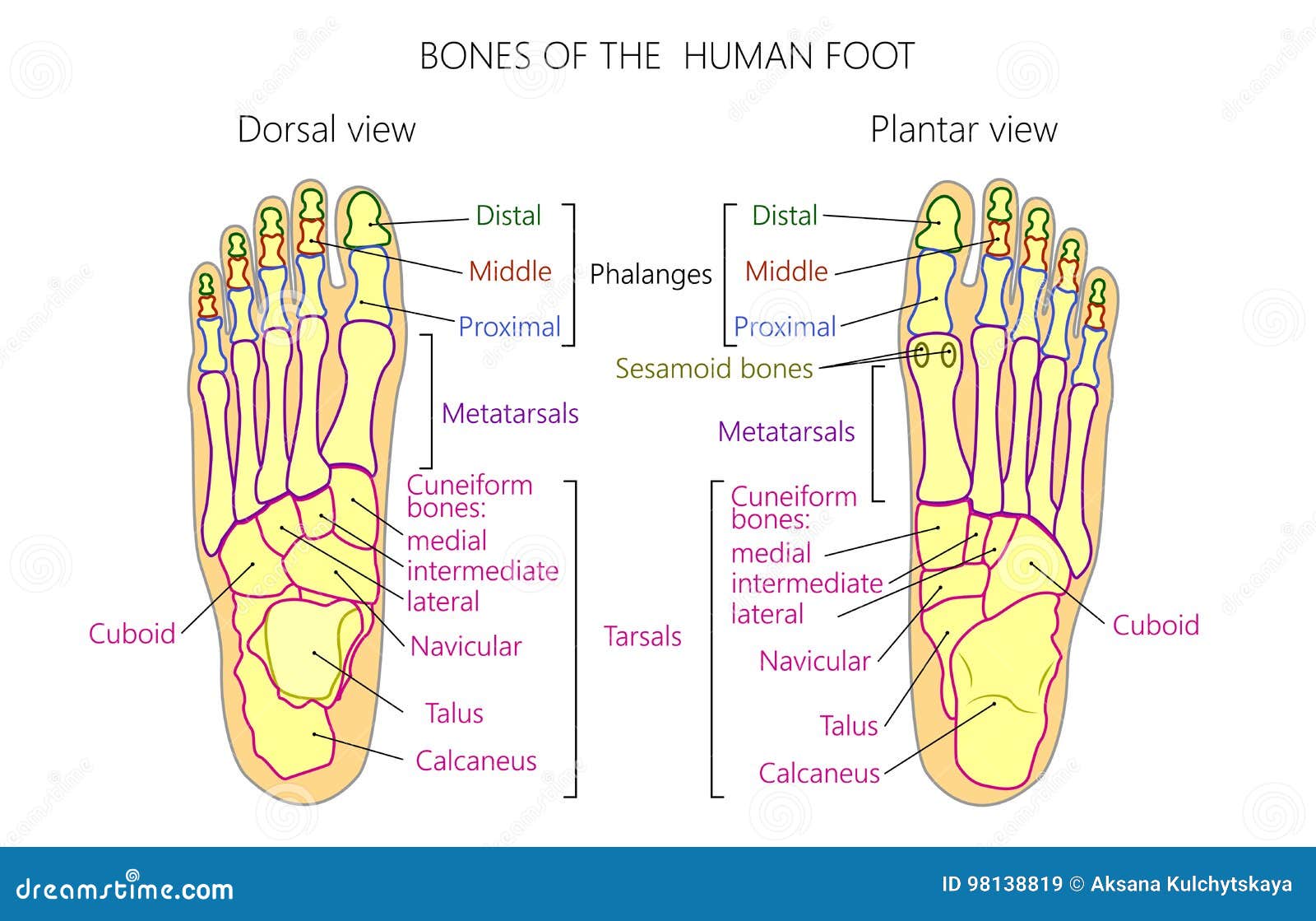
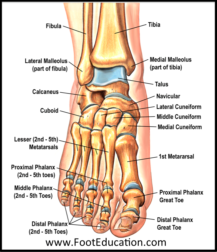
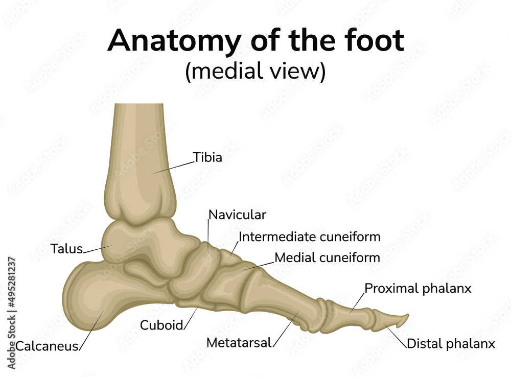
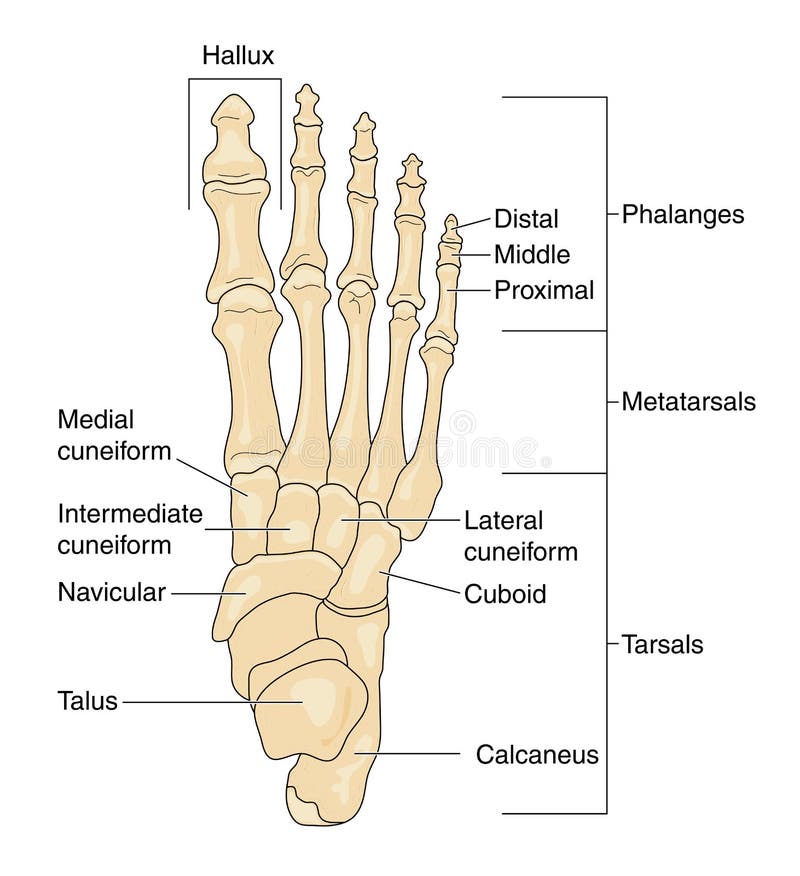
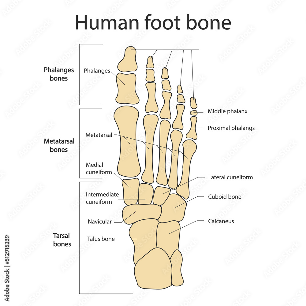
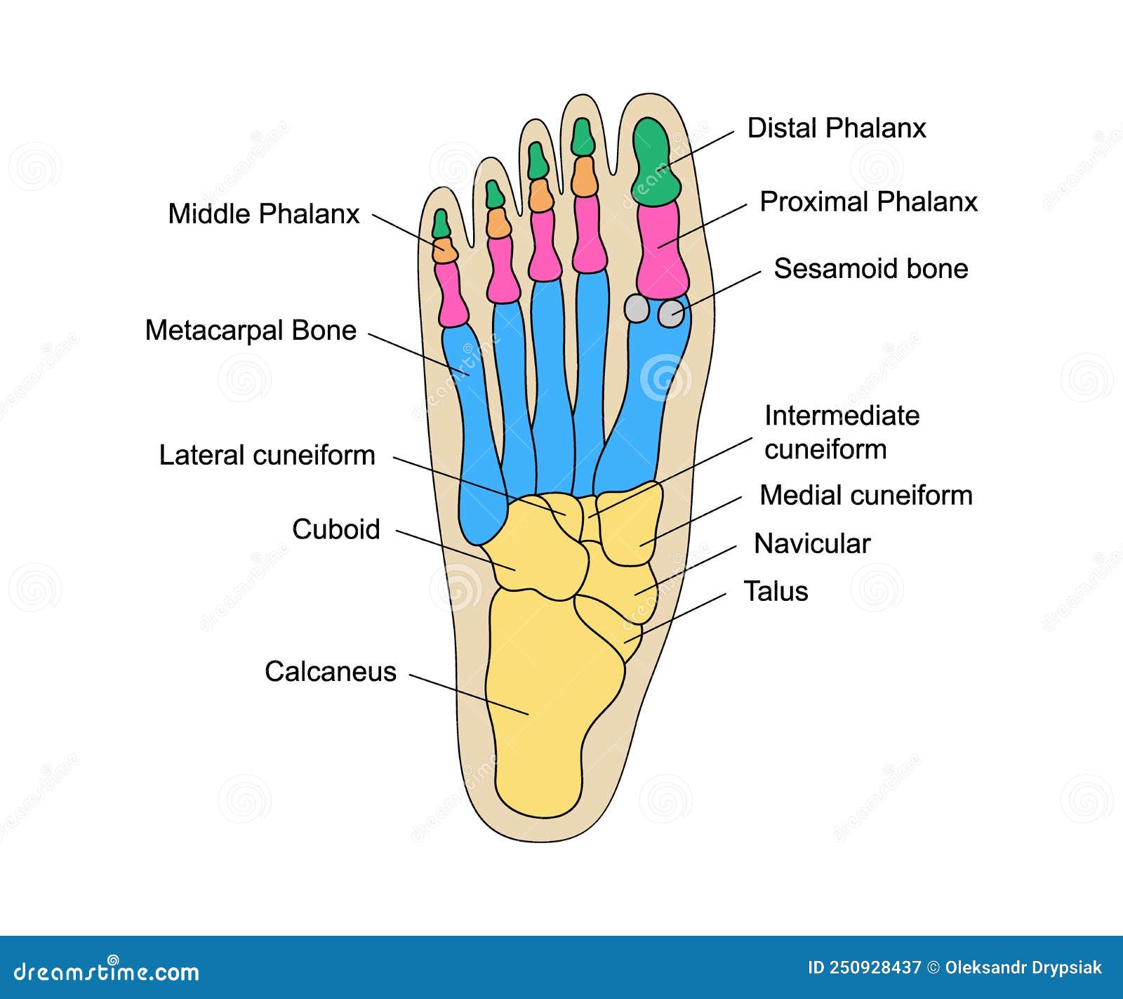
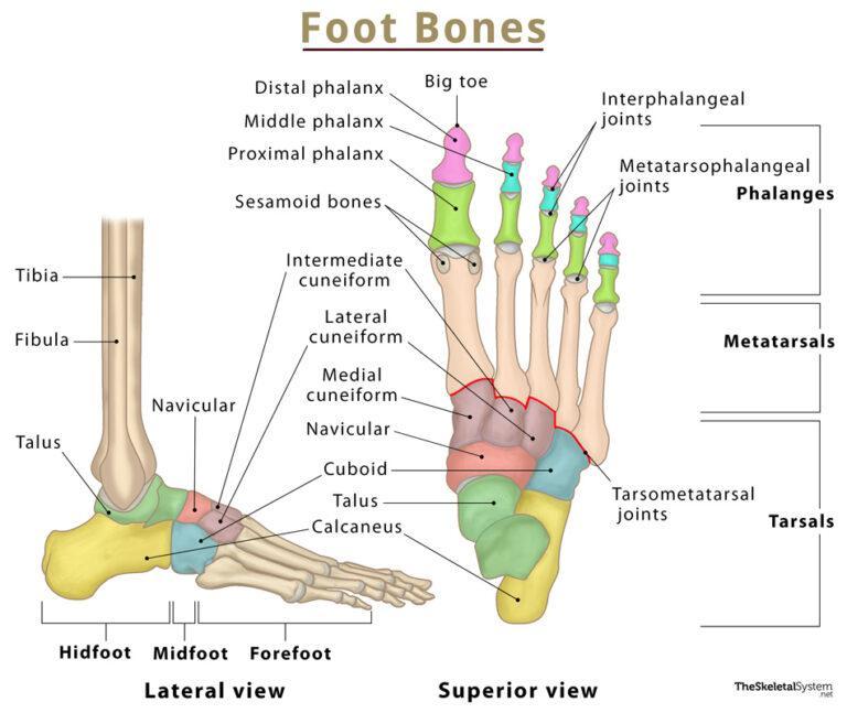
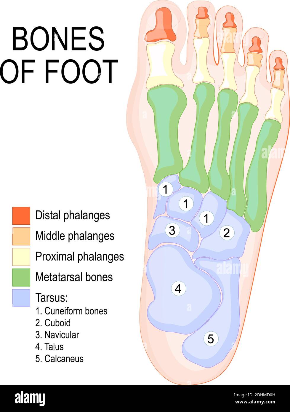
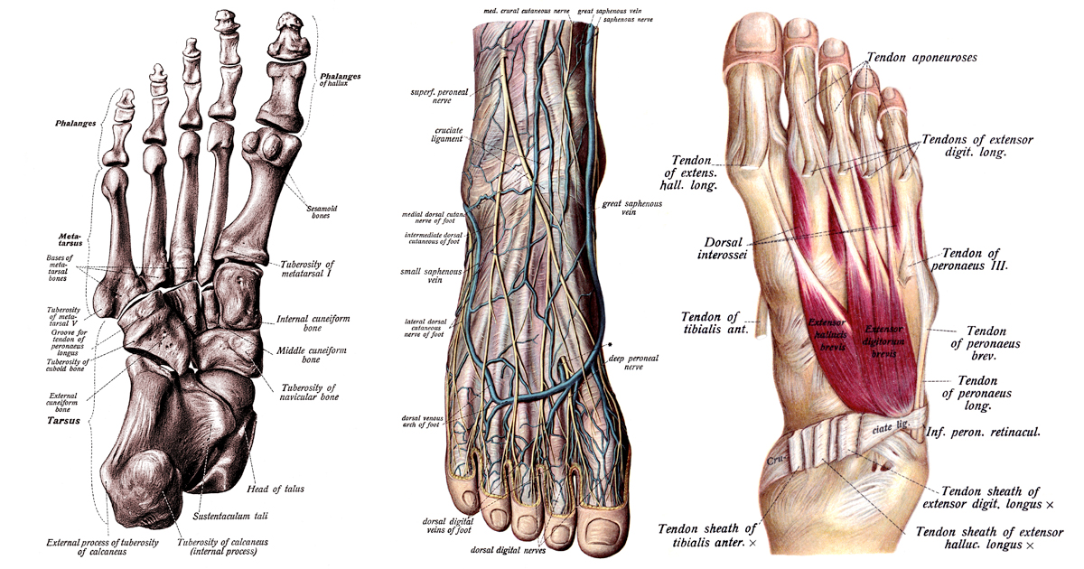
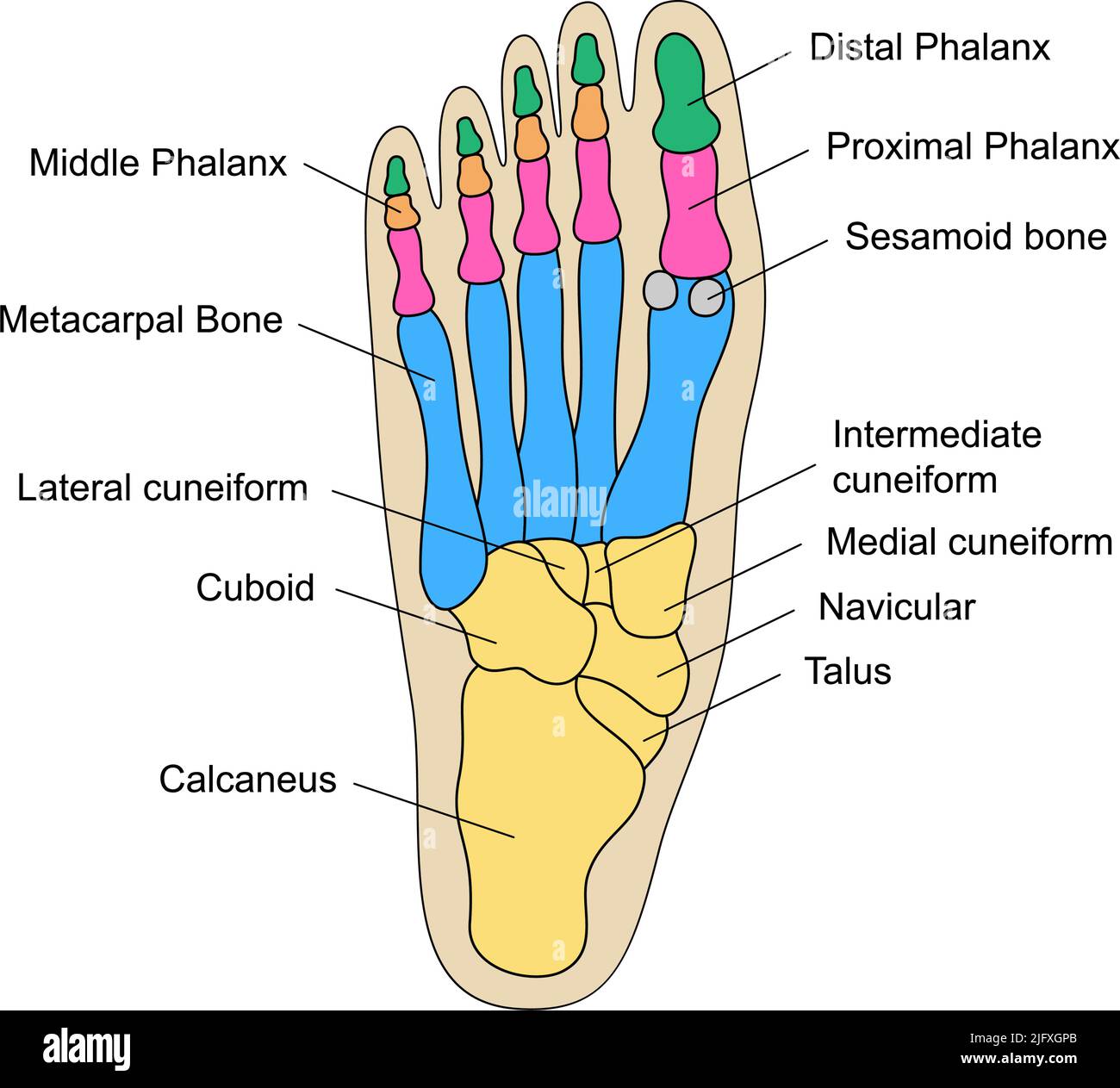
The human foot is an engineering marvel, comprised of 26 bones, almost a quarter of all the bones in your entire body! These bones are divided into three main groups:
-
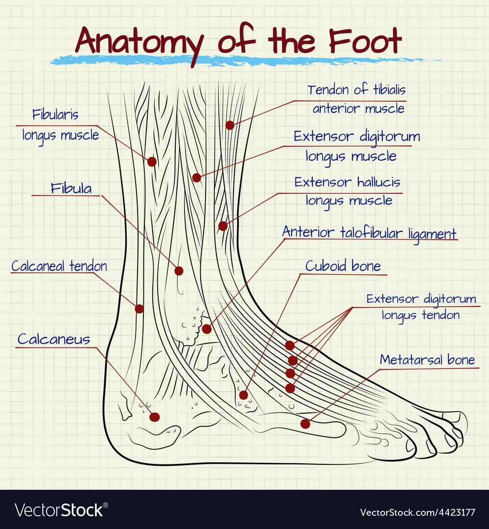
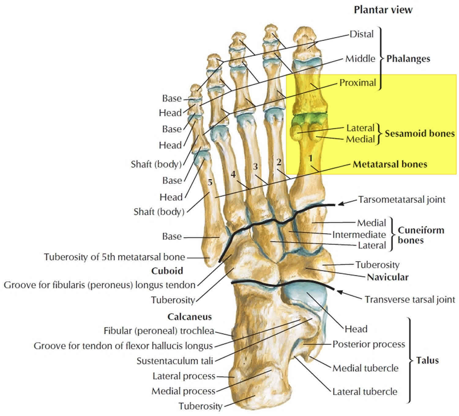
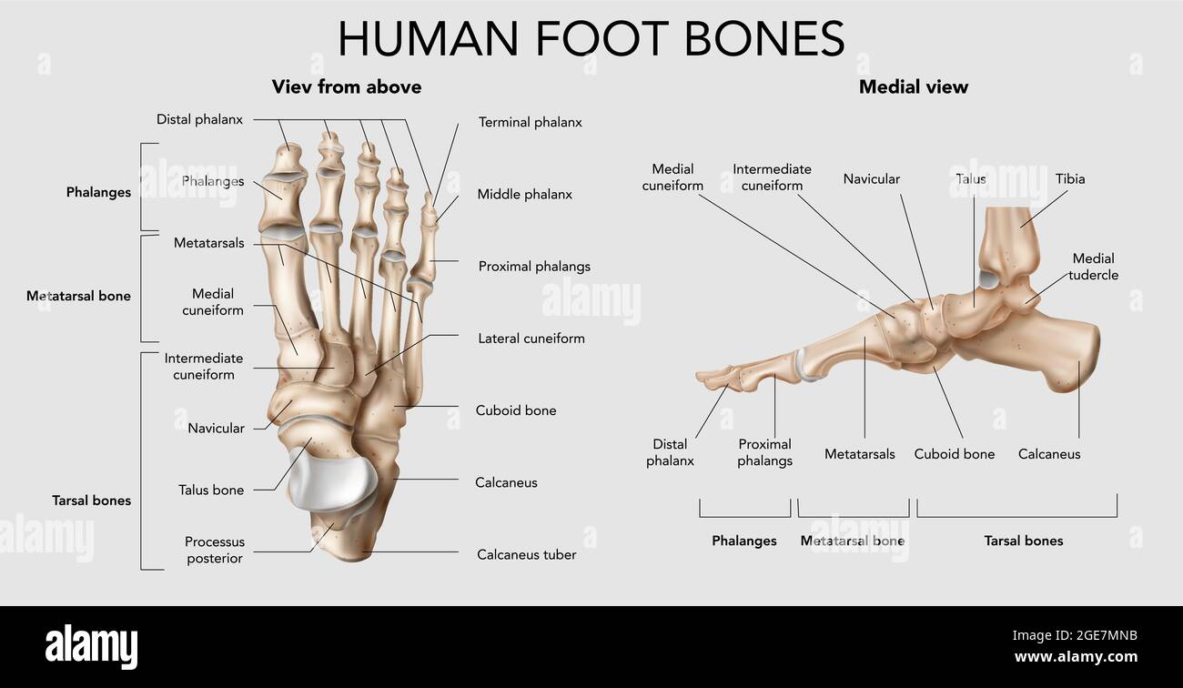
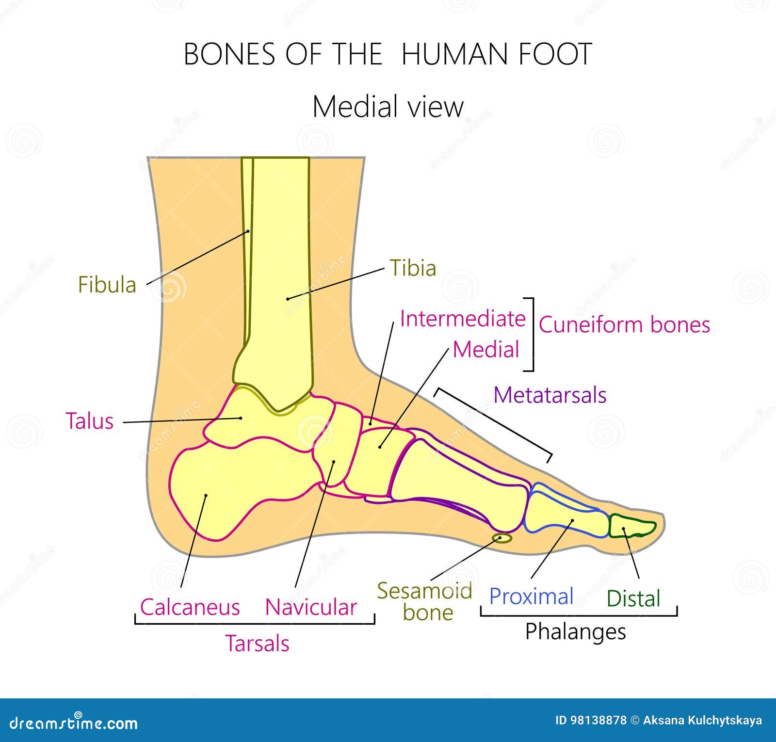
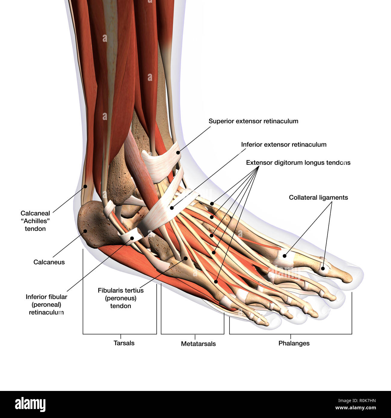
Tarsals: These seven bones form the ankle and the back of the foot. Key tarsals include the talus (which articulates with the tibia and fibula of the lower leg), the calcaneus (heel bone), the navicular, the cuboid, and the three cuneiform bones. Examining a foot anatomy image clearly shows how these bones interlock to provide stability and flexibility.
-
Metatarsals: These five long bones form the arch of the foot and connect the tarsals to the toes. They are numbered 1-5, starting with the big toe side. A detailed foot anatomy image highlights how these bones bear weight and facilitate movement.
-
Phalanges: These are the bones of the toes. The big toe (hallux) has two phalanges (proximal and distal), while the other four toes each have three (proximal, middle, and distal). A foot anatomy image will demonstrate the different lengths and positions of these bones, contributing to the foot's unique shape and function.
Muscles and Tendons: The Movers and Shakers - Foot Anatomy Image
Multiple muscles, both intrinsic (originating and inserting within the foot) and extrinsic (originating in the lower leg), work together to move the foot and ankle. These muscles attach to the bones via tendons, strong cords of tissue.
-
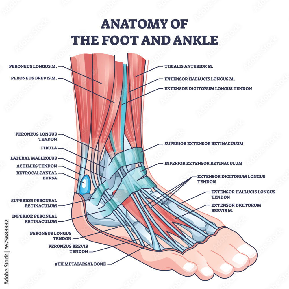
Extrinsic Muscles: These powerful muscles, like the tibialis anterior (dorsiflexion - lifting the foot up), gastrocnemius and soleus (plantarflexion - pointing the toes down), and peroneals (eversion - turning the sole of the foot outward), are crucial for locomotion. Foot anatomy image reveals their origin in the lower leg and their long tendons that reach into the foot.
-
Intrinsic Muscles: These smaller muscles within the foot provide fine motor control and help maintain the arch. They are responsible for movements like curling the toes and providing stability. A detailed foot anatomy image shows their complex arrangement within the sole of the foot.
Ligaments: The Connectors and Stabilizers - Foot Anatomy Image
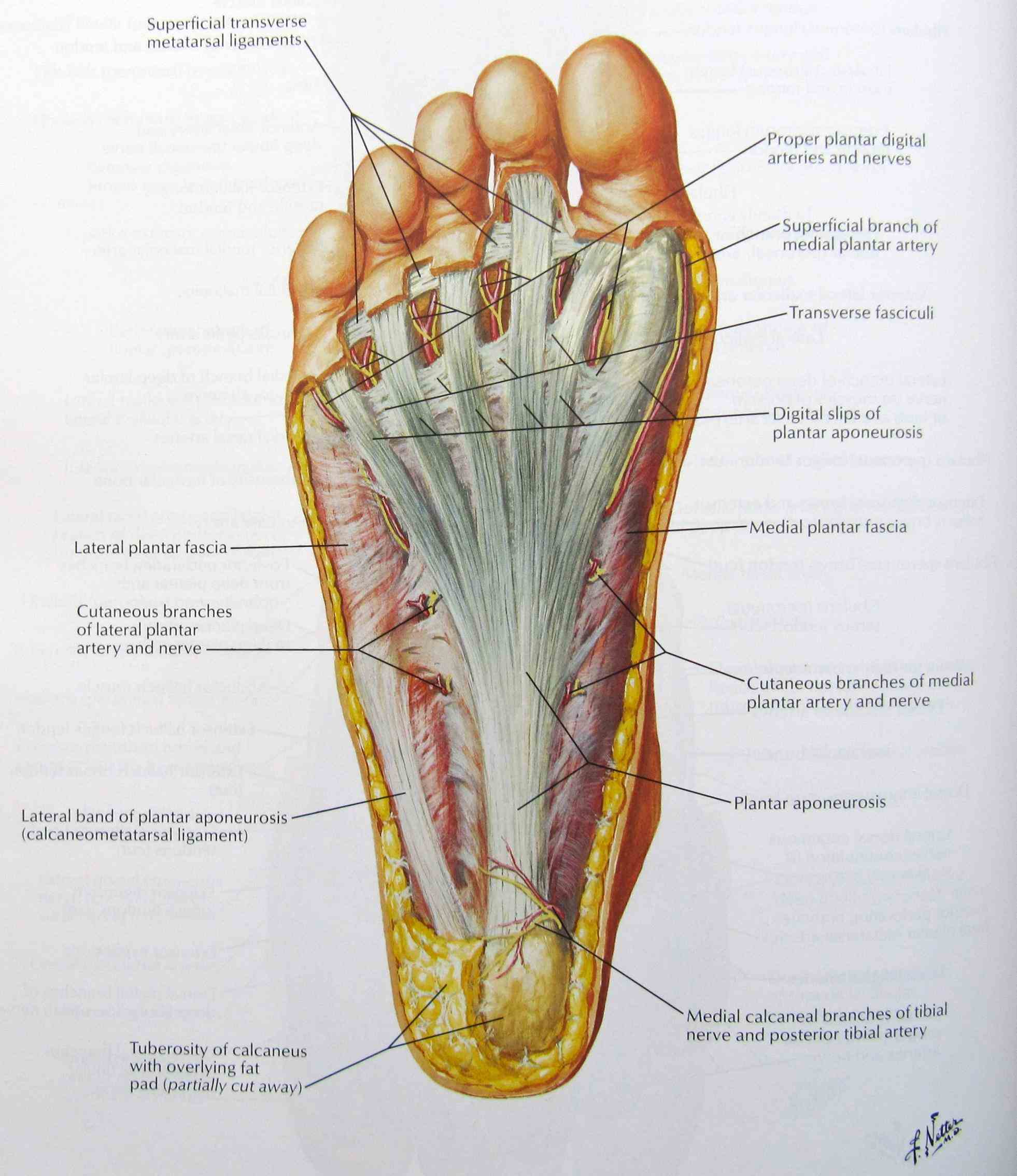
Ligaments are strong, fibrous tissues that connect bones to each other, providing stability and preventing excessive movement. The foot and ankle have numerous ligaments, including the deltoid ligament (on the medial side of the ankle), the lateral ligaments (on the lateral side of the ankle), and the plantar fascia (a thick band of tissue that runs along the bottom of the foot). Understanding a foot anatomy image is essential to appreciate how these ligaments support the bones and prevent injuries.
Nerves and Blood Vessels: Vital Pathways - Foot Anatomy Image
Nerves transmit signals from the brain to the muscles, allowing for movement and sensation. Blood vessels supply the foot with oxygen and nutrients. Major nerves in the foot include the tibial nerve, the peroneal nerve, and the sural nerve. Careful examination of a foot anatomy image illustrates how these nerves and vessels run alongside the bones and muscles, ensuring proper function and health.
Common Foot Problems and the Importance of Foot Anatomy Image Knowledge
Understanding foot anatomy can help prevent and manage common foot problems:
-
Plantar Fasciitis: Inflammation of the plantar fascia, causing heel pain. A foot anatomy image helps visualize the location of the plantar fascia and the source of the pain.
-
Bunions: A bony bump that forms at the base of the big toe. A foot anatomy image can illustrate the misalignment of the bones in a bunion.
-
Sprains: Injuries to ligaments, often caused by sudden twisting or turning. A foot anatomy image shows which ligaments are most vulnerable to sprains.
-
Fractures: Breaks in the bones of the foot, caused by trauma or stress. A foot anatomy image is essential for identifying the location and severity of the fracture.
Taking Care of Your Feet: Proactive Steps
- Wear supportive shoes: Choose shoes that fit well and provide adequate arch support.
- Stretch regularly: Stretching the calf muscles and plantar fascia can help prevent injuries.
- Maintain a healthy weight: Excess weight puts extra stress on the feet.
- See a podiatrist: Consult a podiatrist for any foot pain or problems.
The Power of Informational Style in Foot Health
By moving beyond fleeting trends and focusing on a detailed understanding of foot anatomy image, we can make informed decisions about foot care and injury prevention. This knowledge empowers us to take control of our foot health and enjoy a more active and comfortable life. Educating yourself on foot anatomy image is the first step toward a healthier, happier you.
Trending this Week: Foot Anatomy Image Search Volume
This week has seen a surge in online searches for "foot anatomy image" due to increased awareness campaigns about foot health and the rise in fitness activities. People are actively seeking information to better understand their bodies and prevent injuries.
Question and Answer About Foot Anatomy Image
Q: What is the most important bone in the foot shown in a foot anatomy image?
A: While all bones in the foot are important, the talus is arguably the most crucial. It's the bone that connects the foot to the lower leg and bears a significant amount of weight.
Q: Can a foot anatomy image help me diagnose my foot pain?
A: A foot anatomy image can provide a visual understanding of the structures in your foot, but it's not a substitute for a professional diagnosis. Consult a doctor or podiatrist for an accurate assessment of your pain.
Q: Where can I find a good foot anatomy image?
A: Many medical websites and online resources offer detailed foot anatomy images. Search for reputable sources, such as those from universities or medical journals.
Q: Why is understanding foot anatomy image important for athletes?
A: Athletes need to understand their foot anatomy image to optimize performance, prevent injuries, and recover effectively from training. Knowing the muscles, ligaments, and bones involved in movement can help athletes tailor their training and footwear to their specific needs.
Q: What is the Plantar Fascia?
A: The plantar fascia is a thick band of tissue that runs along the bottom of your foot, from your heel to your toes. The plantar fascia supports the arch of your foot and acts as a shock absorber.
Who is the Celebrities and why did they talk about foot problem?
- Usain Bolt: Is a retired Jamaican sprinter, widely considered the greatest sprinter of all time. He holds the world records in the 100 metres, 200 metres, and 4 x 100 metres relay. Sprinters often face foot problems due to the high impact and repetitive stress placed on their feet during training and competition. Problems like plantar fasciitis, stress fractures, and tendonitis are common. Usain Bolt has discussed foot problems he faced during his career, including foot injuries that impacted his training and performance. These issues are a reality for many high-performance athletes. Understanding foot anatomy image becomes critical in managing and preventing such injuries, making it relevant in discussions about athletic performance and health.
In summary, understanding foot anatomy image is crucial for everyone, especially athletes, for preventing injuries and maintaining foot health. Knowing the bones, muscles, ligaments, and nerves allows for informed decisions about foot care. Common problems like plantar fasciitis, bunions, and sprains can be better understood with a detailed foot anatomy image. Question and Answer confirm that while images aid understanding, professional diagnosis is essential. Keywords: foot anatomy image, plantar fasciitis, bunions, sprains, foot pain, foot health, foot bones, foot muscles, foot ligaments, talus, metatarsals, phalanges, tibialis anterior, gastrocnemius, soleus.
Foot And Ankle Anatomy Explained By Surgeon Andy Hughes Foot Anatomy 2 700x523 Human Foot Anatomy Hi Res Stock Photography And Images Alamy Anatomy Of Human Foot With Labels R0K7HN Infographic Diagram Of Human Foot Bone Anatomy System Lateral View 3D 1000 F 220913545 BSofD1IwNhjImuvZe35yVOlelsscxJ8z Anatomy Of The Foot Medical Art Library Foot Anatomy Foot Anatomy Illustration Shown Is A Medial View Of The Bones Of The 1000 F 495281237 5Z9sgRJRktPE9aK7F1j8K07UPpfDYroc Anatomy Of Foot And Ankle With Labeled Medical Location Outline Diagram 1000 F 675688382 TaJStsPPTb9YYQujEogDgzn2mOw2zVo9 Foot Anatomy Illustration Stock Image F029 5278 Science Photo F0295278 Foot Anatomy, Illustration
Foot Bones Anatomy Of The Skeletal System Of The Human Legs And Feet 1000 F 512915239 BApmPAccaplmFzANH9jbEQ40kbUor5WK Plantar Anatomy Of The Foot TrialQuest Inc FA C 0231 Library Medium Right Foot Anatomy Solved The Figure Illustrates A Medial View Of FA C 0290 Library Medium Foot Anatomy Stock Illustration CartoonDealer Com 30725125 Foot Anatomy Medically Accurate Illustration 58829505 Download Human Foot Anatomy Png 8 Wallpapers Com Human Foot Anatomy Png 8 Xibauabm4vutiox6 Foot Bones Anatomy Realistic Foot Bones Anatomy Infographic Composition With Top And Side Views Of Footstep Skeleton With Text Vector Illustration 2GE7MNB The Anatomy Of The Foot Karen S Whimsy The Anatomy Of The Foot Life Drawing Reference Female Pose Reference Anatomy Reference Photo Bc8a0daaff18b21ee8c726ef3b673867
Bones Of The Foot Labeled Royalty Free Stock Photo Image 8616465 Bones Foot Labeled 8616465 Premium Photo Detailed 3D Representation Of Foot Anatomy Detailed 3d Representation Foot Anatomy 85952 4482 Diagram Of Foot Anatomy New Hampshire Podiatrist Foot Anatomy 691202530 Human Foot Bones Anatomy With Descriptions Educational Diagram Of Human Foot Bones Anatomy Descriptions Colored Leg Base Parts Structure Educational Diagram Internal Organ Illustration Talus 250928437 DIAGRAM Diagram Of Foot Structure MYDIAGRAM ONLINE Foot And Toe Anatomy Plantar View Foot Description Drawings Bones Facts Britannica Bones Foot Tarsal Bones Talus Metatarsal Calcaneus Medial Foot Anatomy Anatomy Bones Human Foot Medial View Vector Illustration Leg Denominations Side 98138878 DIAGRAM Diagram Of Foot Structure MYDIAGRAM ONLINE Structure Of The Human Foot Vector 4423177
Human Foot Bones Anatomy With Descriptions Educational Diagram Of Human Foot Bones Anatomy With Descriptions Educational Diagram Of Internal Organ Illustration Calcaneus Tarsals Navicular Cuboid Foot Parts 2JFXGPB Regions Of Foot Anatomy Anatomical Landmarks On The Dorsum Of The Left Foot Showing Surface Anatomy Of EDB Muscle.ppmPremium Photo A Detailed Foot Diagram With The Various Body Parts And Detailed Foot Diagram With Various Body Parts Anatomical Themes Highlighted Their Correct Positions Your Feet Labeled From Top Bottom 865659 23990 Bones Of Foot Human Anatomy The Diagram Shows The Placement And Names Bones Of Foot Human Anatomy The Diagram Shows The Placement And Names Of All Bones Of Foot 2DHMD0H Foot Medical Diagram Figure 1 Bones Of The Foot And Ankle Diagram Of Foot Anatomy Foot Bones Of Leg And Foot Form Part Appendicular Skeleton That Supports Many Muscles Lower Limbs These Work Together Foot Bones Names Anatomy Structure Labeled Diagrams Foot Bones Labeled Diagram 768x647
Premium Photo Detailed Foot Anatomy Illustration With Care Techniques Detailed Foot Anatomy Illustration With Care Techniques Educational Health Design Medical Posters Print Media 416256 15461 Foot Anatomy Plantar View Anatomy Bones Human Foot Dorsal Plantar View Vector Illustration Leg Denominations Views 98138819 Anatomical Foot Body Reflexology Connection Chart WordLayouts Anatomical Foot Body Reflexology Connection 12 86eqqgbx2