Last update images today Decoding Feet: Anatomy Images Function Amp Care
Decoding Feet: Anatomy Images, Function & Care
Introduction: Unveiling the Complex World of Foot Anatomy Images
This week, we're diving deep into the fascinating world of foot anatomy. Often overlooked, our feet are intricate structures that play a crucial role in our daily lives. From supporting our weight to enabling movement, their health is paramount. This article will explore foot anatomy images, common foot problems, and how to maintain optimal foot health. Whether you're an athlete, someone experiencing foot pain, or simply curious about the human body, this guide is for you.
Target Audience: Athletes, individuals experiencing foot pain, healthcare professionals, students studying anatomy, and anyone interested in learning more about foot health.
Understanding the Bones: Images of Foot Anatomy and Skeletal Structure
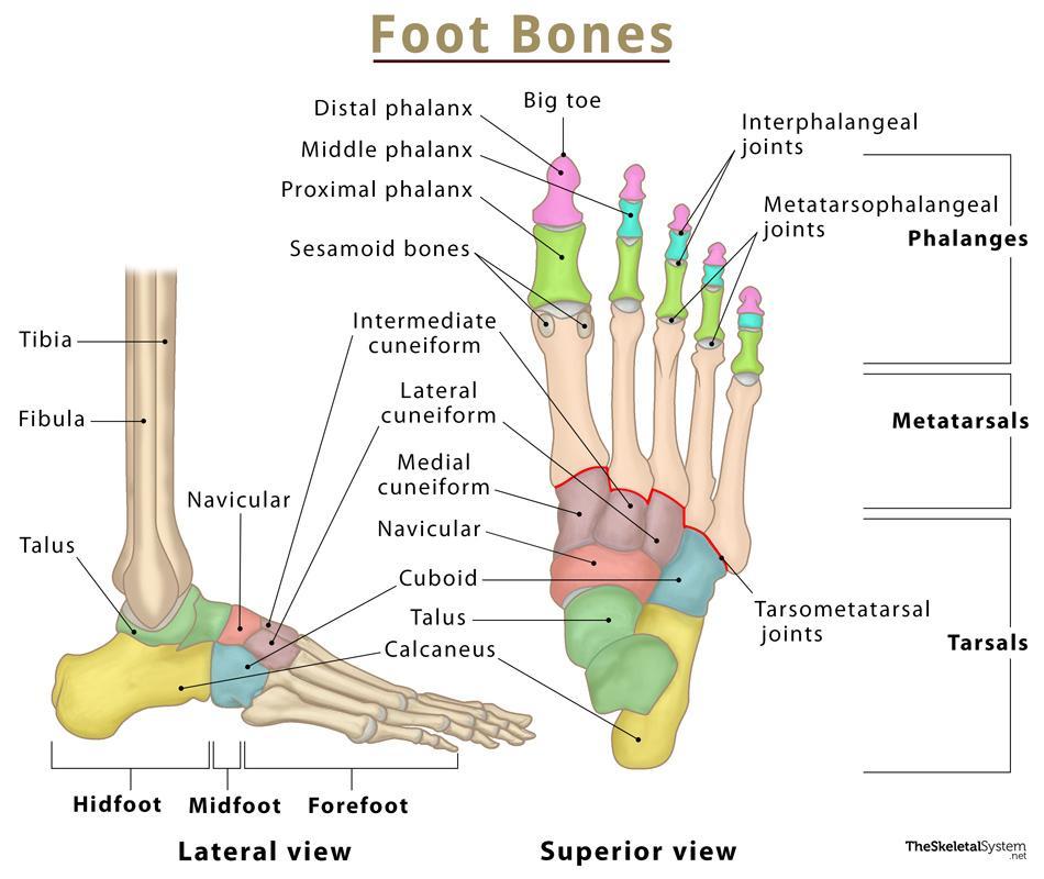
The human foot is a marvel of engineering, comprised of 26 bones, 33 joints, and over 100 muscles, tendons, and ligaments. Understanding the skeletal structure, which you can easily visualize using images of foot anatomy, is fundamental.
-
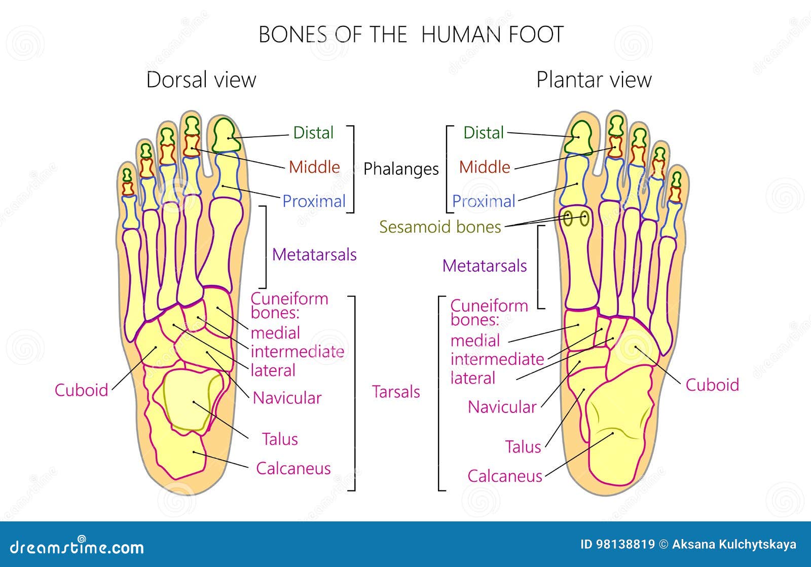
The Foot's Divisions: The foot is divided into three main parts:
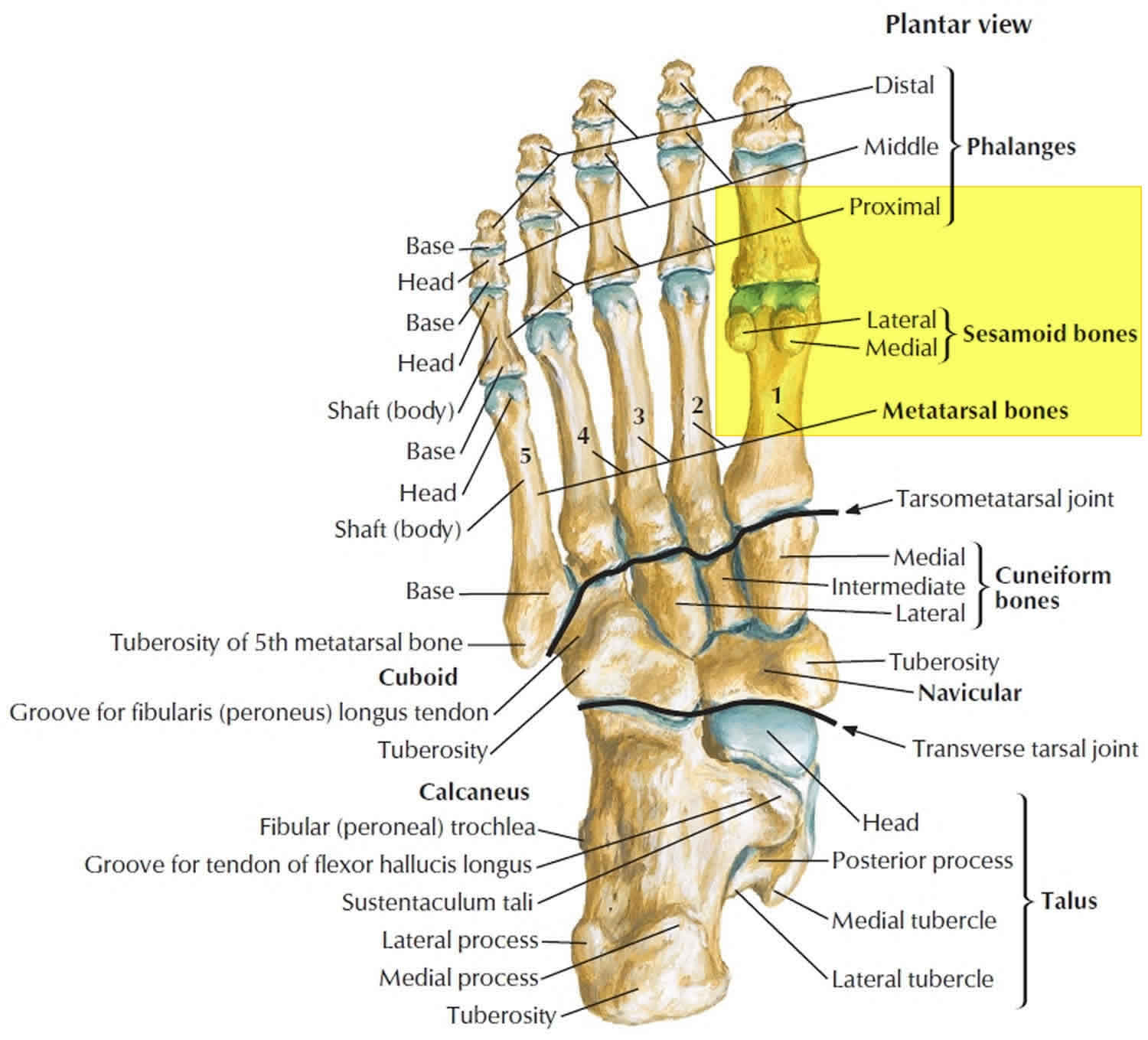
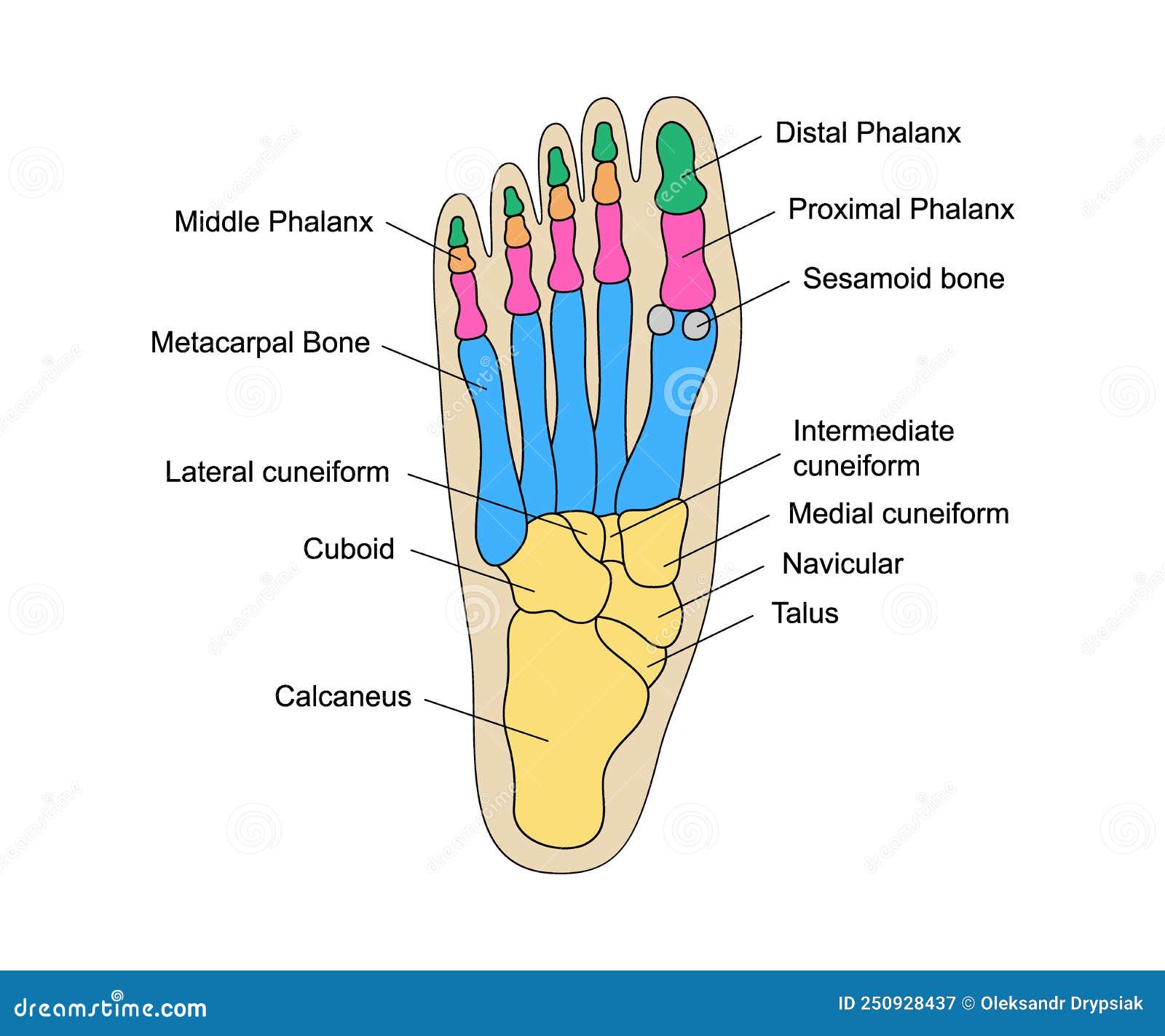
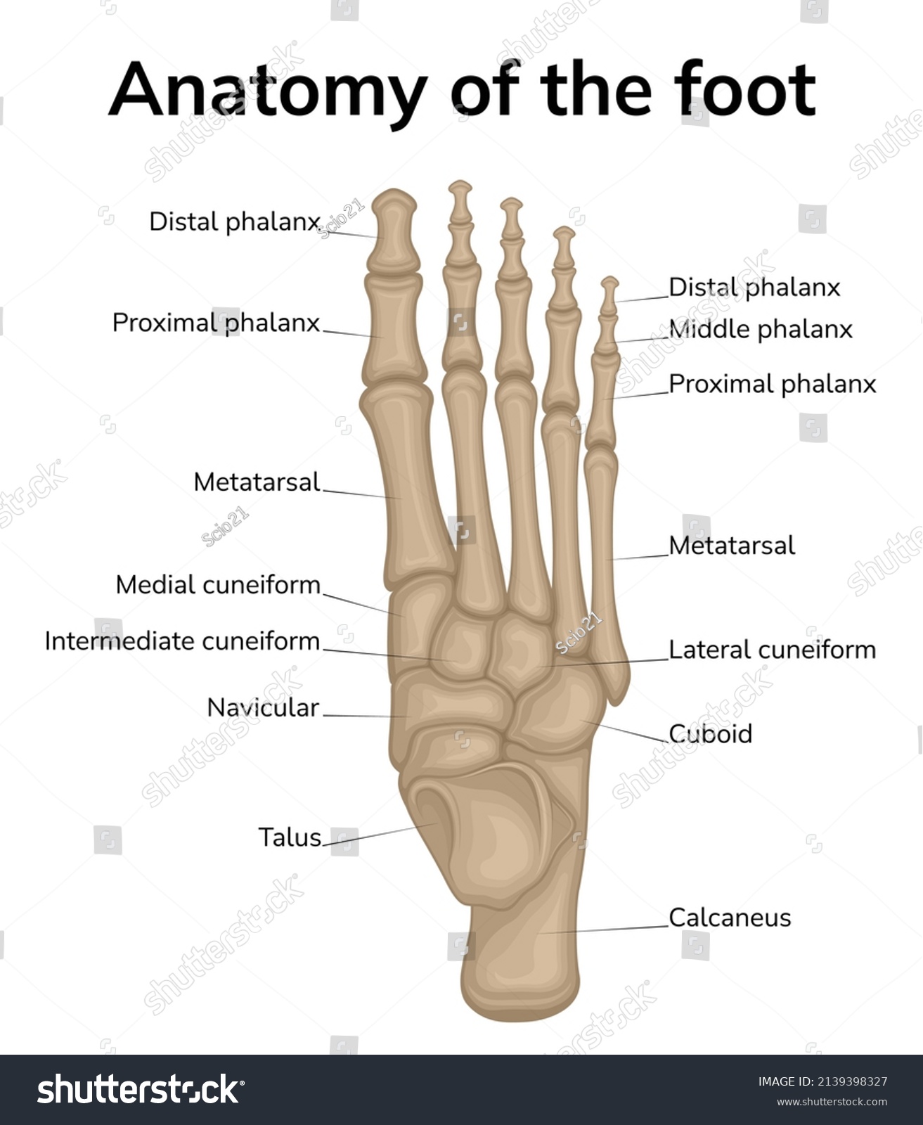
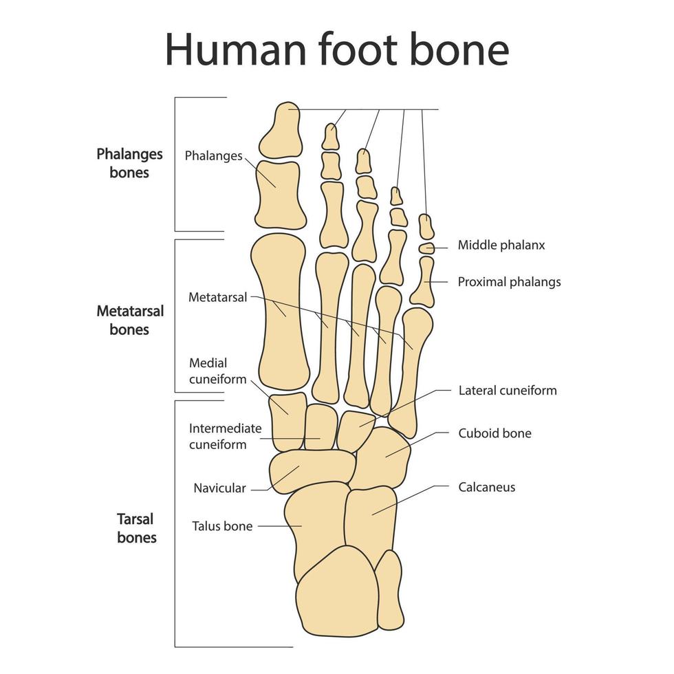
- Forefoot: Contains the metatarsals (long bones) and phalanges (toe bones). Images of foot anatomy clearly show how the metatarsals connect to the toes, allowing for flexibility and balance.
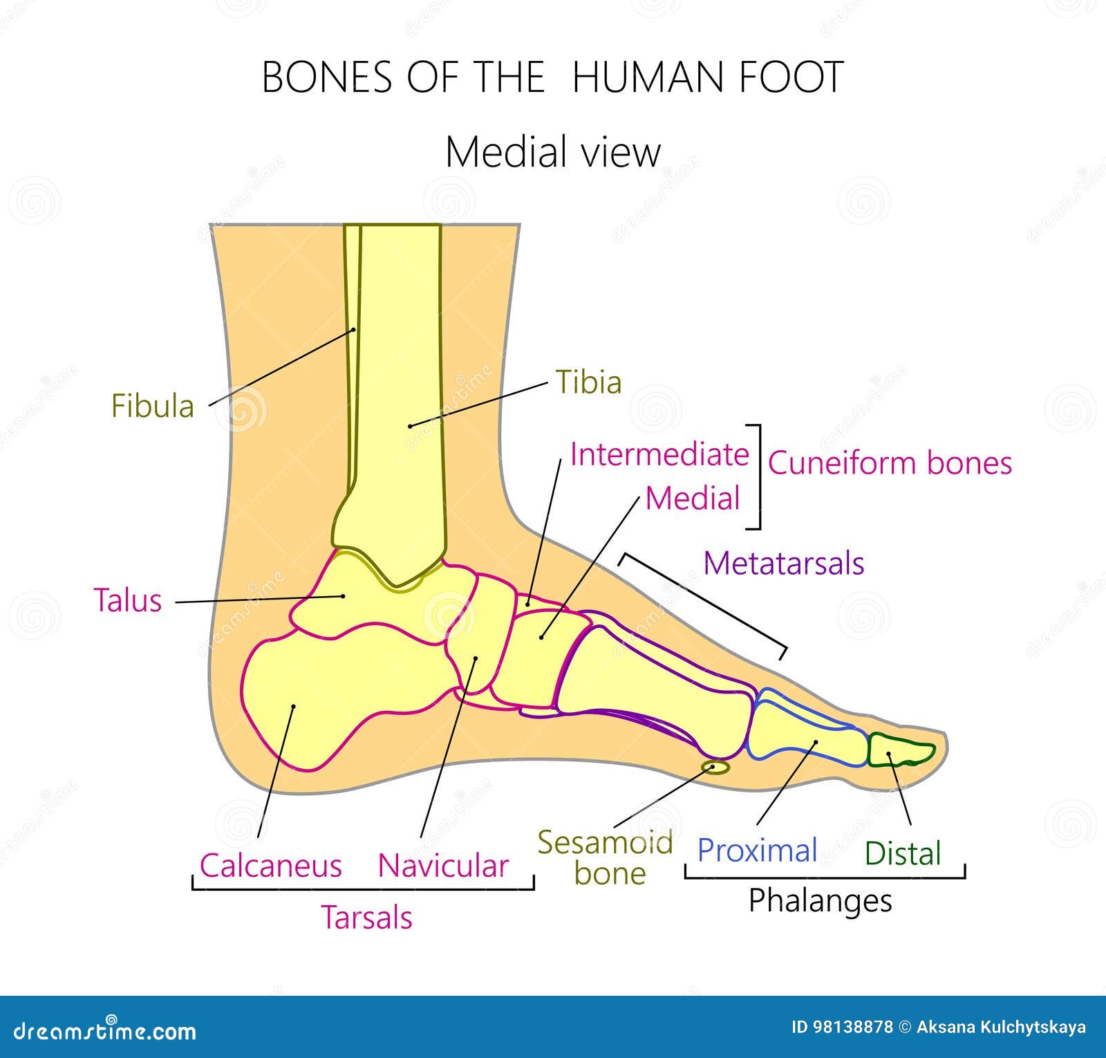
- Midfoot: Houses the cuneiforms, cuboid, and navicular bones. These bones form the arch of the foot, providing support and shock absorption. Viewing images of foot anatomy highlighting the midfoot's structure is essential for understanding arch support.
- Hindfoot: Composed of the talus (ankle bone) and calcaneus (heel bone). The talus articulates with the tibia and fibula of the lower leg, forming the ankle joint. Images of foot anatomy illustrate the critical role the hindfoot plays in weight distribution and stability.
-
Key Bones and Their Functions:
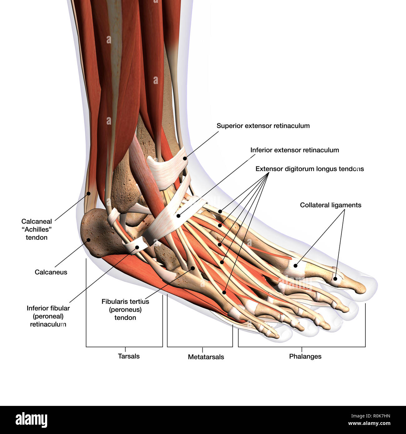
- Calcaneus (Heel Bone): The largest bone in the foot, it bears the brunt of our weight and provides attachment points for the Achilles tendon. Images of foot anatomy will show the prominence of the calcaneus.
- Talus (Ankle Bone): Sits atop the calcaneus and connects the foot to the lower leg. It's crucial for ankle movement. You can find images of foot anatomy detailing its complex articulation.
- Metatarsals: Five long bones that connect the ankle to the toes. They play a key role in weight distribution during walking and running. Images of foot anatomy showing the metatarsals help understand stress fractures.
- Phalanges (Toe Bones): Each toe has three phalanges (proximal, middle, and distal), except for the big toe, which has only two. They provide flexibility and balance. Images of foot anatomy will highlight the different sizes and shapes of the phalanges.
Muscles, Tendons, and Ligaments: Foot Anatomy Images of Soft Tissues
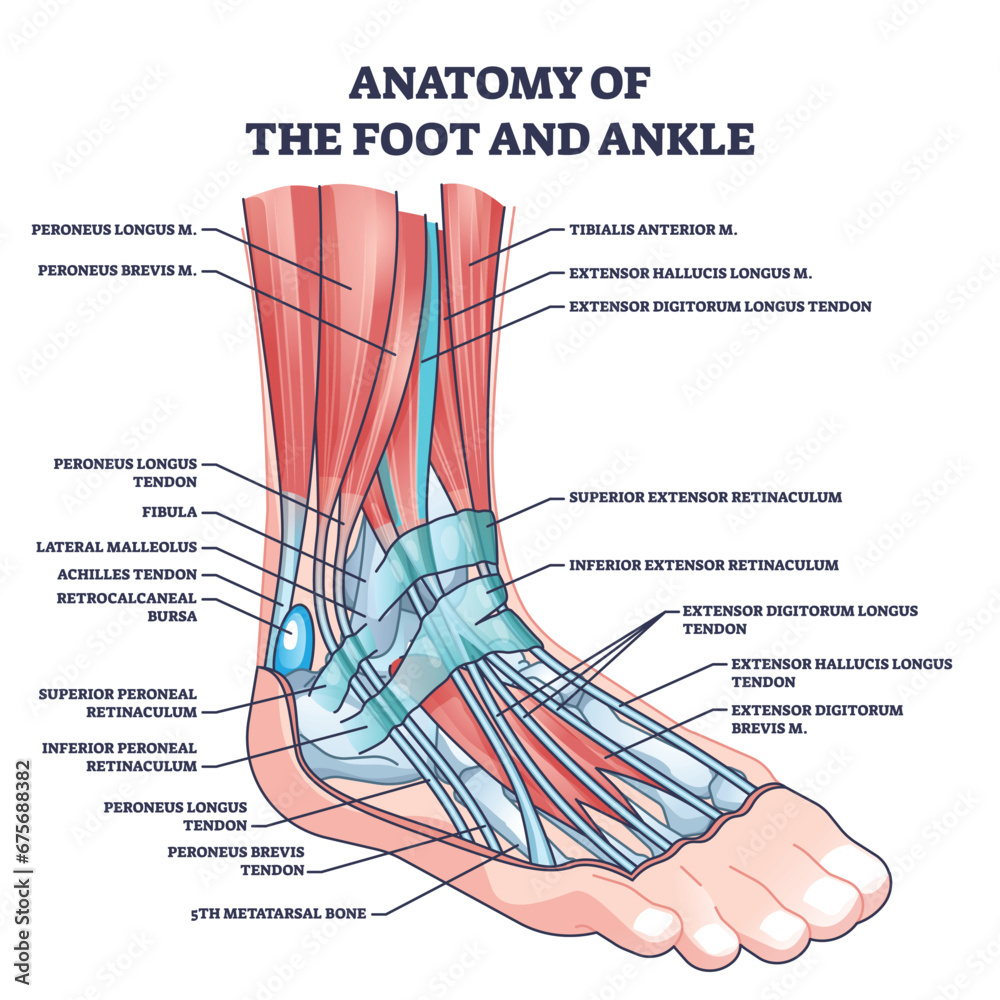
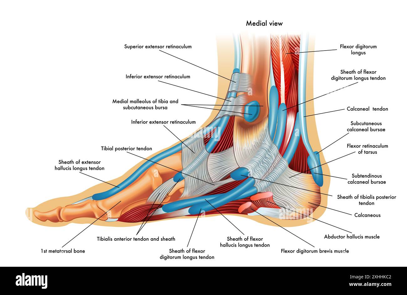
Beyond bones, the soft tissues of the foot are equally important. Images of foot anatomy help visualize these complex structures.
-
Muscles: The foot contains both intrinsic (within the foot) and extrinsic (originating in the leg) muscles.
- Intrinsic Muscles: Control fine movements of the toes and help maintain the arch.
- Extrinsic Muscles: Responsible for dorsiflexion (lifting the foot), plantarflexion (pointing the foot), inversion (turning the sole inward), and eversion (turning the sole outward).
-
Tendons: Connect muscles to bones. The Achilles tendon, the largest tendon in the body, connects the calf muscles to the calcaneus. Images of foot anatomy illustrate how tendons facilitate movement.
-
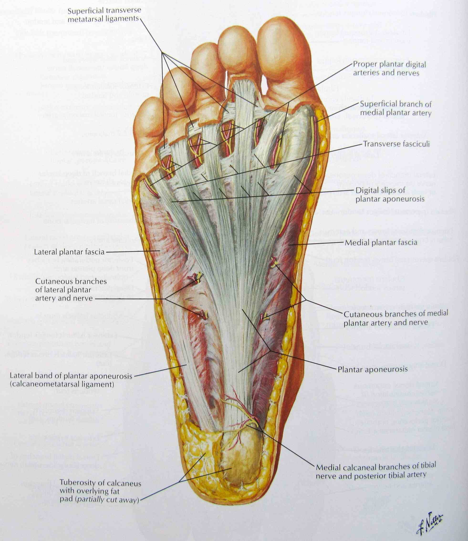
Ligaments: Connect bone to bone, providing stability to the joints. The plantar fascia, a thick band of tissue on the bottom of the foot, supports the arch and absorbs shock. Images of foot anatomy highlight the importance of ligaments in joint stability.
Common Foot Problems: A Visual Guide with Images of Foot Anatomy
Understanding the anatomy helps in identifying and addressing common foot problems. Images of foot anatomy coupled with symptom descriptions can be incredibly helpful.
- Plantar Fasciitis: Inflammation of the plantar fascia, causing heel pain. Images of foot anatomy will show the location of the plantar fascia and where the inflammation typically occurs.
- Bunions: A bony bump that forms on the joint at the base of the big toe. Images of foot anatomy clearly demonstrate the misalignment of the joint.
- Hammertoe: A deformity where a toe bends abnormally at the middle joint. Images of foot anatomy show the characteristic bend in the toe.
- Athlete's Foot: A fungal infection that causes itching, burning, and cracking of the skin on the feet. Images of foot anatomy would illustrate how fungal infections can look on the skin.
- Ankle Sprains: Occur when ligaments are stretched or torn, often due to sudden twisting or injury. Images of foot anatomy highlight the ligaments involved in ankle stability.
- Stress Fractures: Small cracks in the bones, often caused by repetitive stress. Images of foot anatomy show where stress fractures commonly occur in the metatarsals.
Maintaining Foot Health: Tips & Recommendations Using Foot Anatomy Images
Taking care of your feet is crucial for overall well-being. Understanding images of foot anatomy helps in making informed decisions about foot care.
- Proper Footwear: Wear shoes that fit well, provide adequate support, and have good shock absorption.
- Regular Stretching: Stretch your feet and ankles daily to improve flexibility and prevent injuries.
- Weight Management: Maintaining a healthy weight reduces stress on your feet.
- Good Hygiene: Keep your feet clean and dry to prevent fungal infections.
- Orthotics: Consider using orthotics (shoe inserts) for added support and cushioning.
- Professional Care: See a podiatrist (foot specialist) for any persistent foot pain or problems. Using images of foot anatomy, the podiatrist can better explain your condition and treatment options.
Q & A: Addressing Your Foot Anatomy Questions with Images of Foot Anatomy
Here are some common questions related to foot anatomy, answered with a focus on the visual understanding that images of foot anatomy provide.
Q: What is the arch of the foot and why is it important?
A: The arch is the curved structure formed by the bones and ligaments of the midfoot. Using images of foot anatomy, you can see that it acts as a shock absorber, distributing weight evenly and providing flexibility.
Q: What causes plantar fasciitis?
A: Plantar fasciitis is caused by inflammation of the plantar fascia, a thick band of tissue on the bottom of the foot. Overuse, improper footwear, and tight calf muscles can contribute. Images of foot anatomy help illustrate the location and function of the plantar fascia, making it easier to understand how strain leads to inflammation.
Q: How can I prevent foot problems?
A: Choose supportive footwear, stretch regularly, maintain a healthy weight, and practice good foot hygiene. If you experience persistent pain or discomfort, consult a podiatrist. Understanding your foot anatomy using images is the first step in preventative care.
Q: What is the best way to treat a sprained ankle?
A: The RICE method (Rest, Ice, Compression, Elevation) is generally recommended for treating ankle sprains. Over-the-counter pain relievers can also help. If the sprain is severe, seek medical attention. Images of foot anatomy can show the affected ligaments and the severity of the tear or sprain.
Celebrities Who've Talked About Foot Health:
While direct anecdotes about foot anatomy are rare, celebrities often discuss foot-related issues like wearing high heels or dealing with injuries. Let's take a look at one celebrity who's been open about foot health challenges:
Victoria Beckham:
Who is Victoria Beckham? Victoria Beckham, born April 17, 1974, is an English singer, fashion designer, and businesswoman. She rose to fame as a member of the Spice Girls, one of the best-selling girl groups of all time. After the Spice Girls disbanded, Beckham pursued a solo music career before transitioning into the fashion industry. She launched her own fashion label in 2008, which has become known for its sophisticated and elegant designs. Victoria Beckham is married to former footballer David Beckham, and they have four children together. She is considered a style icon and a prominent figure in the fashion world.
Victoria Beckham, known for her love of high heels, has reportedly suffered from bunions and other foot problems. She's even been photographed wearing flats on occasion, a testament to the impact of footwear on foot health! While she hasn't directly discussed "images of foot anatomy," her experiences underscore the importance of proper foot care, especially when wearing fashionable but potentially problematic footwear.
Conclusion: Step Forward with Knowledge of Images of Foot Anatomy
Our feet are essential for our mobility and overall well-being. By understanding foot anatomy images, common foot problems, and preventive measures, we can take better care of our feet and maintain an active, healthy lifestyle. Remember to consult a podiatrist for any persistent foot pain or concerns.
Keywords: images of foot anatomy, foot anatomy, foot pain, plantar fasciitis, bunions, hammertoe, athlete's foot, ankle sprain, stress fracture, foot health, podiatrist, bones of the foot, muscles of the foot, ligaments of the foot, foot care.
Summary Q&A: This article explores the importance of understanding foot anatomy through images, covering the skeletal structure, muscles, common problems, and maintenance tips. Key questions addressed include the function of the foot arch, causes of plantar fasciitis, prevention of foot problems, and treatment for ankle sprains, all aided by visualizing images of foot anatomy.
Right Foot Anatomy Solved The Figure Illustrates A Medial View Of FA C 0290 Library Medium Anatomy Foot Diagram Diagram Of Foot Foot Anatomy Illustration Whit Annotations Stock Photo Alamy Foot Anatomy Illustration Whit Annotations 2XHHKC2 Regions Of Foot Anatomy Anatomical Landmarks On The Dorsum Of The Left Foot Showing Surface Anatomy Of EDB Muscle.ppmThe Anatomy Of The Foot Karen S Whimsy The Anatomy Of The Foot Anatomy Of The Foot And Ankle By Podiatrist Denver CO Elite Foot Anatomy Foot Ankle 549x600 Premium Photo A Detailed Foot Diagram With The Various Body Parts And Detailed Foot Diagram With Various Body Parts Anatomical Themes Highlighted Their Correct Positions Your Feet Labeled From Top Bottom 865659 24037 Premium Vector Human Foot Bones Anatomy Sketch Vector Orthopedic Human Foot Bones Anatomy Sketch Vector Orthopedic Medicine Skeleton Phalanges Ankles 593268 33
Infographic Diagram Of Human Foot Bone Anatomy System Lateral View 3D 1000 F 220913545 BSofD1IwNhjImuvZe35yVOlelsscxJ8z Foot Anatomy Illustration Shown Top View Stock Vector Royalty Free Stock Vector Foot Anatomy Illustration Shown Is A Top View Of The Bones Of The Foot 2139398327 Diagram Of Foot Anatomy Foot Bones Of Leg And Foot Form Part Appendicular Skeleton That Supports Many Muscles Lower Limbs These Work Together Medial Foot Anatomy Anatomy Bones Human Foot Medial View Vector Illustration Leg Denominations Side 98138878 Foot Diagram With Labels Foot Bones Labeled Diagram Plantar Anatomy Of The Foot TrialQuest Inc FA C 0231 Library Medium DIAGRAM Diagram Of Foot Structure MYDIAGRAM ONLINE Foot And Toe Anatomy Plantar View
Foot Anatomy Illustration Stock Vector Illustration Of Phalanges Foot Anatomy Illustration Shown Medial View Bones Foot Foot Anatomy Illustration 244192275 Anatomy Of Human Foot With Labels Stock Photo Alamy Anatomy Of Human Foot With Labels R0K7HN Premium Photo Detailed Foot Anatomy Illustration With Care Techniques Detailed Foot Anatomy Illustration With Care Techniques Educational Health Design Medical Posters Print Media 416256 15461 List 105 Pictures Pictures Of Foot Anatomy Stunning Image951 DIAGRAM Diagram Of Foot Structure MYDIAGRAM ONLINE L F1007 13354.1456241545.1280.1280 Anatomy Of The Foot And Ankle OrthoPaedia Anatomy Bones Foot D04bc4e8b3d4f1abe4b8730015a5d8e3 Human Foot Bones Anatomy With Descriptions Educational Diagram Of Human Foot Bones Anatomy Descriptions Colored Leg Base Parts Structure Educational Diagram Internal Organ Illustration Talus 250928437 Foot Medical Diagram New Hampshire Podiatrist Foot Anatomy 691202530
Anatomical Diagram Of The Sole Of The Foot Showing The Important Anatomical Diagram Of The Sole Of The Foot Showing The Important Landmarks Foot Bones Anatomy Royalty Free Vector Image VectorStock Foot Bones Anatomy Vector 27231314 Anatomy Of Foot And Ankle With Labeled Medical Location Outline Diagram 1000 F 675688382 TaJStsPPTb9YYQujEogDgzn2mOw2zVo9 Premium Photo Detailed 3D Representation Of Foot Anatomy Detailed 3d Representation Foot Anatomy 85952 4482 Foot And Ankle Anatomy Explained By Surgeon Andy Hughes Foot Anatomy 2 768x574 Anatomy Bones Of The Human Foot Dorsal And Plantar View Stock Vector Anatomy Bones Human Foot Dorsal Plantar View Vector Illustration Leg Denominations Views 98138819 Foot Bones Anatomy Of The Skeletal System Of The Human Legs And Feet Foot Bones Anatomy Of The Skeletal System Of The Human Legs And Feet Vector
Anatomy Of The Foot Bones Bones Of The Foot Labeled Human Foot Anatomy Illustration High Res Vector Graphic Getty Images Human Foot Anatomy Illustration