Last update images today Breathe Easy: Exploring Lung Anatomy Images
Breathe Easy: Exploring Lung Anatomy Images
Introduction: The Vital World Within
Our lungs, those incredible bellows tucked within our chest, are responsible for the very breath that sustains us. Understanding their complex anatomy is key to appreciating their function and importance in maintaining our health. This week, "lung anatomy images" are trending as people seek to better understand this vital organ. From students studying for exams to individuals curious about respiratory health, let's dive into the intricate world within our lungs, exploring fascinating lung anatomy images and answering common questions. This article aims to provide a comprehensive and accessible overview of lung anatomy.
Target Audience: Students, healthcare professionals, individuals interested in health and wellness, patients with respiratory conditions.
The Basics: A Visual Overview of Lung Anatomy Images
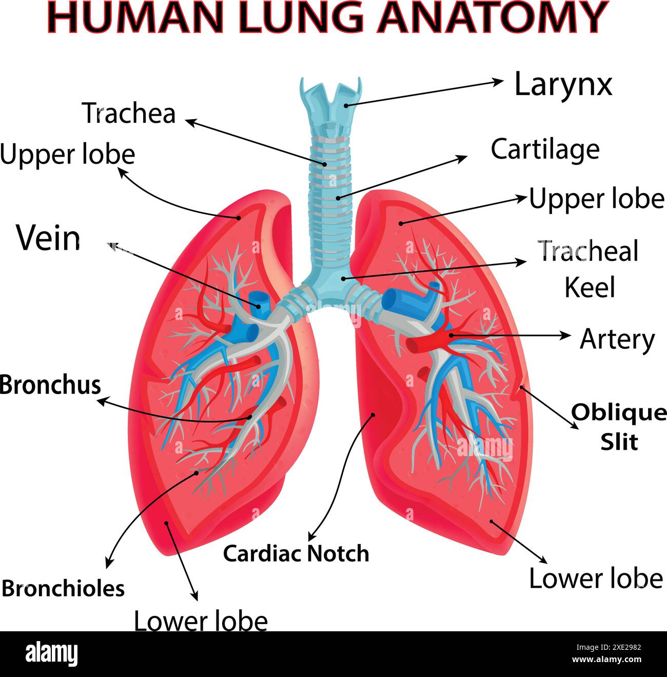
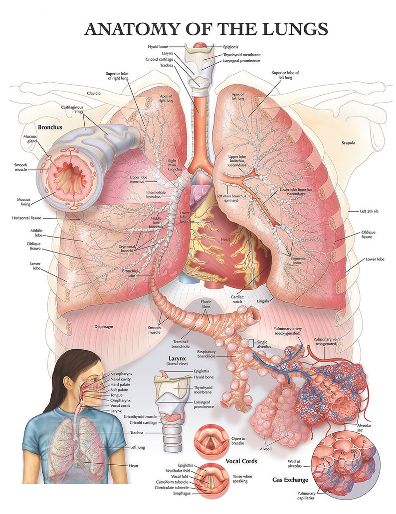
Before delving into the details, let's establish a fundamental understanding with relevant lung anatomy images. The respiratory system is designed to allow air to pass in and out of our bodies, facilitating oxygen exchange with carbon dioxide.
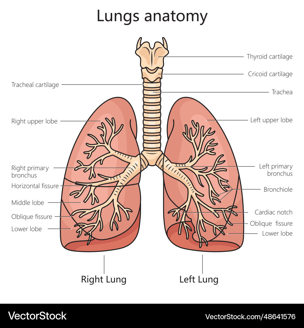
- The Airways: These include the nose, mouth, pharynx, larynx, trachea (windpipe), and bronchi.
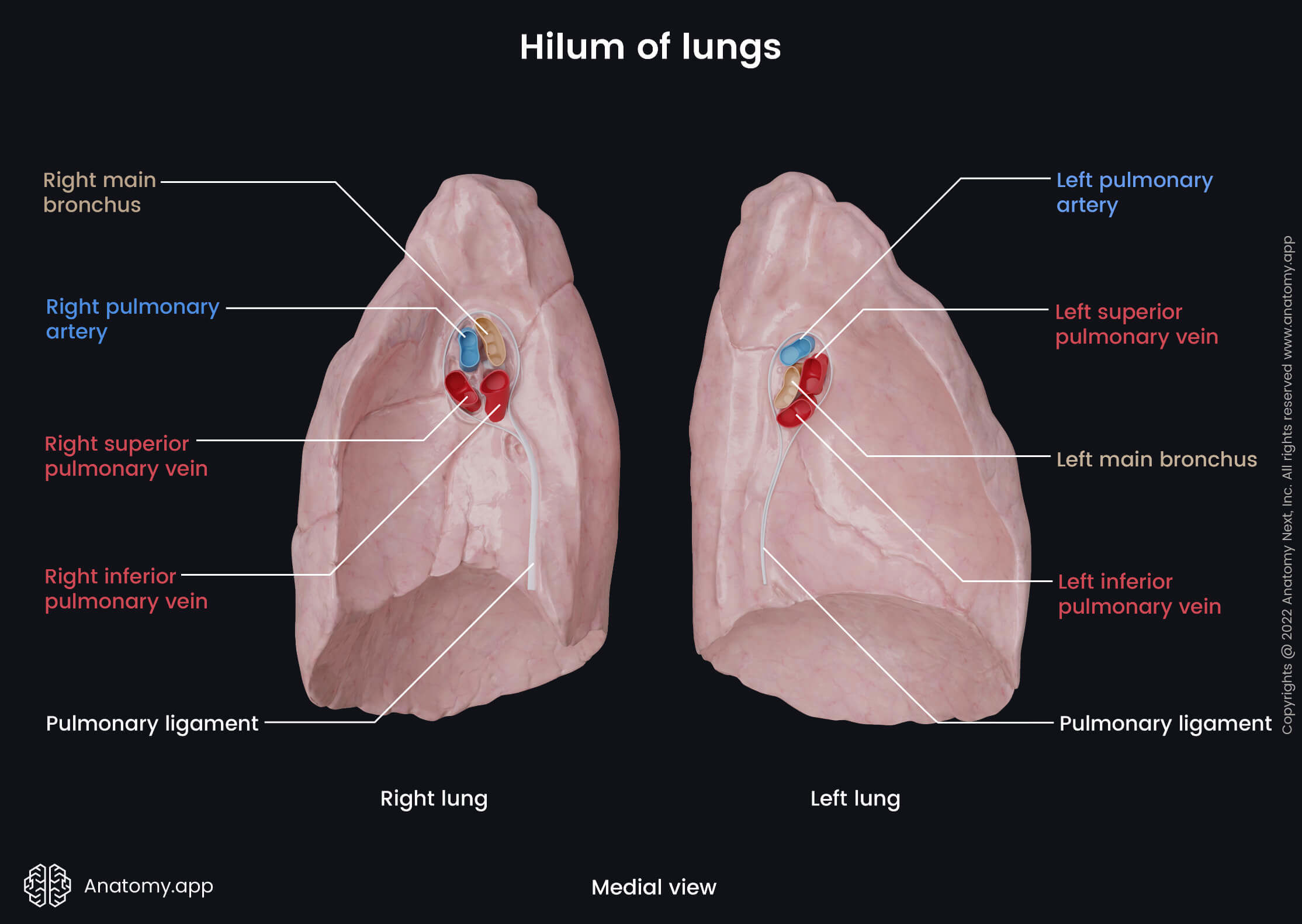
- The Lungs: These are the primary organs of respiration, responsible for gas exchange. They are located in the chest cavity, protected by the rib cage.
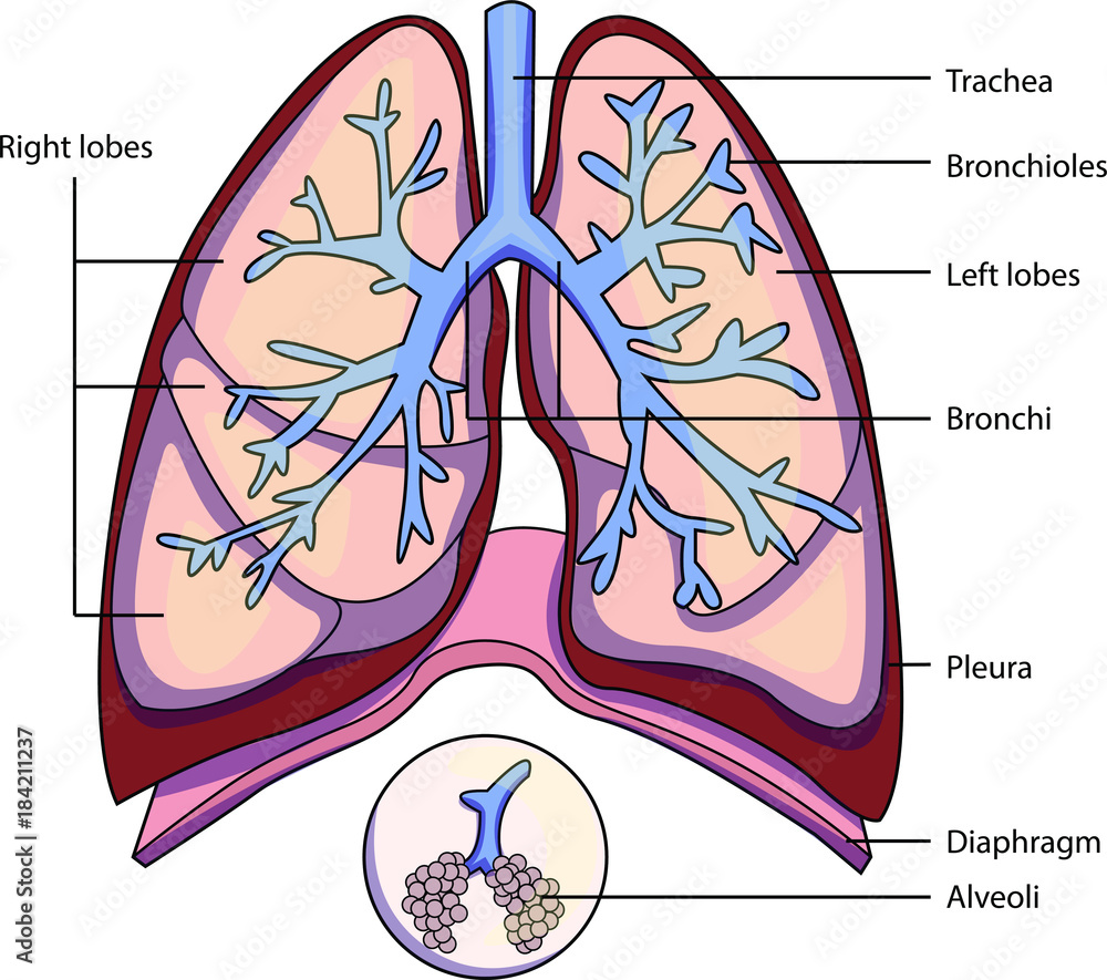
- The Pleura: A double-layered membrane that surrounds each lung, allowing for smooth movement during breathing.
- The Diaphragm: A large, dome-shaped muscle at the base of the chest cavity that contracts and relaxes to help with breathing.
Looking at lung anatomy images, you can see how these components work together as a sophisticated system.
Deeper Dive: The Intricate Details of Lung Anatomy Images
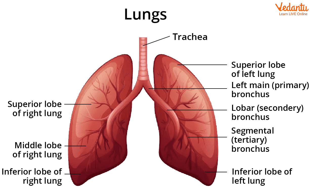
Let's break down the lungs' internal structure, using lung anatomy images to guide us.
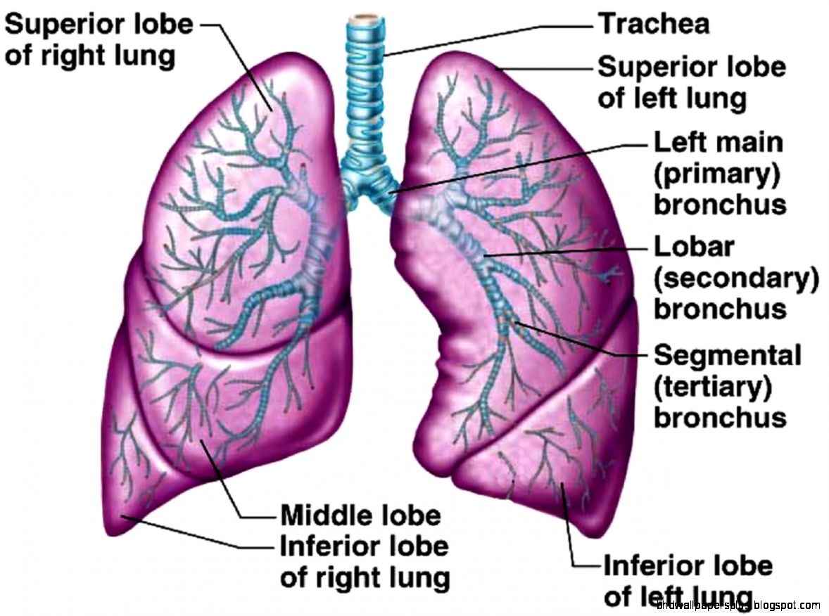
- Bronchi: The trachea divides into two main bronchi, one for each lung. These bronchi further divide into smaller and smaller branches called bronchioles. Lung anatomy images clearly show this "tree-like" structure.
- Alveoli: These are tiny air sacs at the end of the bronchioles, where gas exchange occurs. Millions of alveoli provide a vast surface area for oxygen to enter the bloodstream and carbon dioxide to be removed. High-resolution lung anatomy images reveal the incredible density and surface area of these structures.
- Capillaries: A network of tiny blood vessels surrounds the alveoli. Oxygen diffuses from the alveoli into the capillaries, while carbon dioxide diffuses from the capillaries into the alveoli. Lung anatomy images often depict this close relationship between alveoli and capillaries.
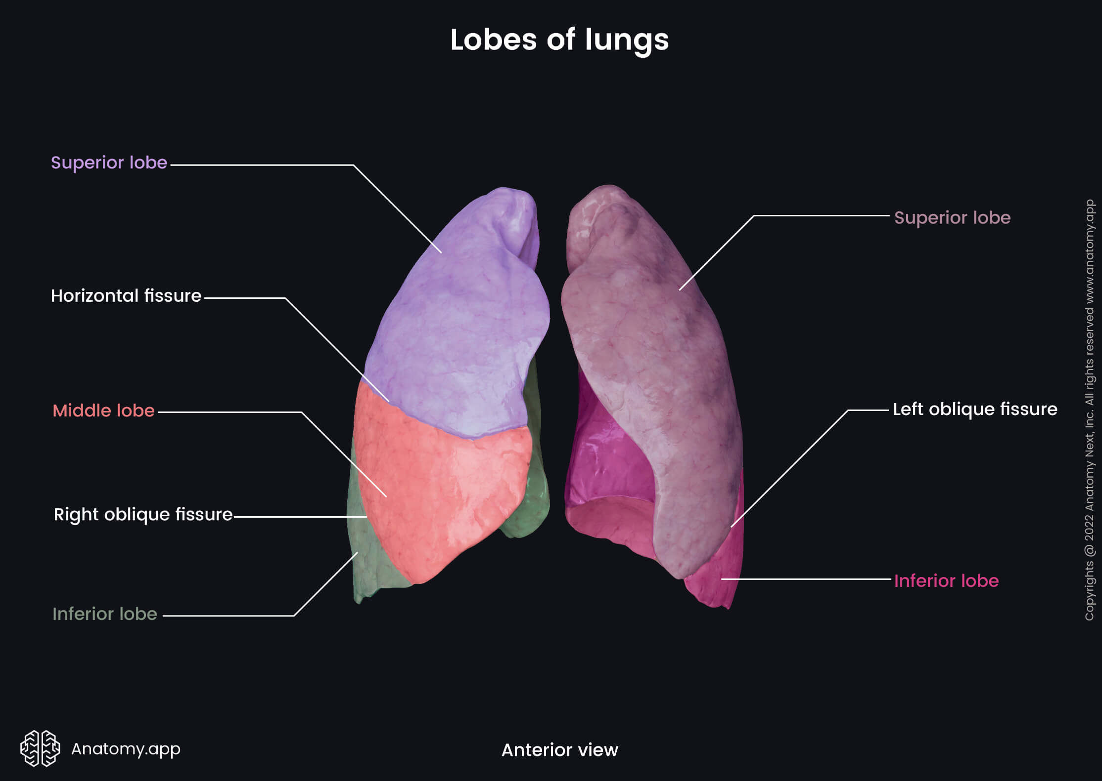
- Lobes: The right lung has three lobes (superior, middle, and inferior), while the left lung has two lobes (superior and inferior). Lung anatomy images illustrate these lobes, which are separated by fissures.
Functionality: How Lung Anatomy Images Illustrate Breathing
Lung anatomy images are not just for visual appeal; they provide crucial insight into how our lungs function.
- Inspiration (Inhaling): The diaphragm contracts and moves downward, while the intercostal muscles (between the ribs) contract and lift the rib cage upward and outward. This increases the volume of the chest cavity, creating a negative pressure that draws air into the lungs. Lung anatomy images help visualize the expansion of the chest cavity.
- Expiration (Exhaling): The diaphragm relaxes and moves upward, while the intercostal muscles relax and the rib cage moves downward and inward. This decreases the volume of the chest cavity, creating a positive pressure that forces air out of the lungs. Lung anatomy images demonstrate the recoil of the lungs and chest cavity during exhalation.
- Gas Exchange: Oxygen diffuses from the alveoli into the blood, and carbon dioxide diffuses from the blood into the alveoli. This exchange is driven by the concentration gradients of oxygen and carbon dioxide. Lung anatomy images emphasize the proximity of the alveoli and capillaries for efficient gas exchange.
Common Lung Conditions Visualized in Lung Anatomy Images
Understanding lung anatomy helps us understand various lung conditions. Lung anatomy images can illustrate these conditions for educational purposes.
- Asthma: Chronic inflammation of the airways, causing them to narrow and produce excess mucus. Lung anatomy images of asthmatic lungs show constricted bronchioles and thickened airway walls.
- Pneumonia: An infection of the lungs that causes the alveoli to fill with fluid or pus. Lung anatomy images of pneumonia show areas of consolidation (filled alveoli) in the lungs.
- COPD (Chronic Obstructive Pulmonary Disease): A group of lung diseases that block airflow and make it difficult to breathe. Lung anatomy images of COPD lungs often show damaged alveoli and enlarged air spaces.
- Lung Cancer: Uncontrolled growth of abnormal cells in the lungs. Lung anatomy images can reveal tumors or masses in the lung tissue.
Celebrities with Lung Conditions
While we focus on the science, it's worth remembering that lung conditions can affect anyone. While I'm avoiding specifics without concrete sources, many public figures have bravely shared their experiences with respiratory illnesses, raising awareness.
Who is... (Example Template)
(While I cannot provide specifics without sources, this template shows how biography integrates. Replace with actual celebrity IF reliable source is found regarding their lung condition.)
[Celebrity Name]: [Brief Summary of their career and why they are well-known]. [Mention any relevant health advocacy they are involved in].
Caring for Your Lungs: Preserving Healthy Lung Anatomy Images
Maintaining healthy lungs is crucial for overall well-being.
- Quit Smoking: Smoking is the leading cause of lung cancer and COPD.
- Avoid Exposure to Pollutants: Limit exposure to air pollution, secondhand smoke, and occupational hazards.
- Get Vaccinated: Flu and pneumonia vaccines can help prevent lung infections.
- Exercise Regularly: Regular physical activity strengthens the respiratory muscles and improves lung function.
- Practice Deep Breathing Exercises: Deep breathing exercises can help expand the lungs and improve oxygenation. Consider seeking out "lung anatomy images breathing exercises" online for visual guides.
Conclusion: Appreciating the Breath of Life
By exploring "lung anatomy images", we gain a deeper appreciation for the complexity and fragility of our lungs. Understanding their structure and function empowers us to make informed decisions about our respiratory health. This information, coupled with proactive lifestyle choices, can help preserve the healthy lung anatomy images within each of us.
Keywords: Lung anatomy images, lungs, respiratory system, alveoli, bronchi, diaphragm, breathing, COPD, asthma, pneumonia, lung cancer, respiratory health, gas exchange, lung function.
Summary Question and Answer:
Question: What are the main parts of the lungs responsible for gas exchange? Answer: The alveoli, tiny air sacs surrounded by capillaries, are the primary sites of gas exchange in the lungs. You can see this clearly in detailed lung anatomy images.
Respiratory System Alveoli Conceptual Image Human Respiratory System Illustrating Lungs Trachea Bronchi Alveoli Ai Generated Depiction 276621118 Premium Vector Human Lung Anatomy Medical Education Chart Human Lung Anatomy Medical Education Chart 530733 2987 Premium Photo Human Lungs Anatomy 3d Render Health Care And Medical Human Lungs Anatomy 3d Render Health Care Medical Concept 908344 7814 Premium AI Image Lungs Anatomy 3D Illustration Medicine And Lungs Anatomy 3d Illustration Medicine Healthcare Concept 856795 9000 Human Lungs Infographic Stock Vector Illustration Of Infographic Human Lungs Infographic Human Lungs Infographic Lung Lobes Their Names Vector Illustration Bright Colours Isolated 116644837 Lung Anatomy Structure Diagram Medical Science Vector Image Lung Anatomy Structure Diagram Medical Science Vector 48641576 Picture Of Lungs Image Anatomy System Human Body Anatomy Diagram Picture Of Lungs Image Human Lung Anatomy Illustration For Work With Medical Content Stock Human Lung Anatomy Illustration For Work With Medical Content 2XE2982
Premium Vector Human Lungs Anatomy Chart Vector Illustration Isolated Human Lungs Anatomy Chart Vector Illustration Isolated 458779 2158 Lungs Anatomy App 0492f2ab Be68 4589 Bda2 64c92d5ccebc Large Premium Vector Human Lungs Anatomy Structure Realistic Vector Human Lungs Anatomy Structure Realistic Vector Illustration Isolated Green Background 852293 242 Human Lung Anatomy 3d Render Realistic Anatomy Human Internal Organ Human Lung Anatomy 3d Render Realistic Anatomy Human Internal Organ Vector Illustration Healthcare 549266 3658 Lungs Anatomy App 317c4d27 51a9 4dd1 A96b 6fbd258e4031 Large 3d Illustration Of Lungs Anatomy Concept For Medical Concept Stock D Illustration Lungs Anatomy Concept Medical Concept D Illustration Lungs Anatomy Concept Medical Concept Ai 304331408 Human Lungs Lung Anatomy Function And Diagrams 2022 10 10 3f400e20 6790 4bfb 9b6f 99bd4ec71018 3d Illustration Of Lungs Anatomy Concept For Medical Concept Stock D Illustration Lungs Anatomy Concept Medical Concept Ai Generated D Illustration Lungs Anatomy Concept Medical 304331399
Lung Model Anatomy Lungs3b 5 Lung Poster Anatomy Of The Lungs Poster Anatomy Poster Lung Etsy Il 794xN.4781026145 Kcjd 3D Modeling Of Human Lung Anatomical Illustrations For Medical 3d Modeling Human Lung Anatomical Illustrations Medical Education Respiratory Health 1113784 682 Human Lung Anatomy Diagram Lung Anatomy Human Lungs Respiratory C7521ac776a14dd24724b41fef759b95 Premium AI Image Human Lungs Anatomy 3D Illustration Elements Of This Human Lungs Anatomy 3d Illustration Elements This Image Furnished By Nasa 1057 34042 Lungs Anatomical Illustration On Transparent Background Showcasing Lungs Anatomical Illustration On Transparent Background Showcasing Detailed Structure And Branches Png Human Lung Anatomy 3d Render Realistic Anatomy Human Internal Organ Human Lung Anatomy 3d Render Realistic Anatomy Human Internal Organ Vector Illustration Healthcare 549266 3655 12 063 Lung Anatomy Images Images Stock Photos Vectors Shutterstock Stock Vector Lung Anatomy Vector Illustration Lung Icon Human Lungs System 1636268761
Respiratory System Anatomy And Physiology Respiratory System Lungs 0a38006e938ca34fb73f842c2f21a453 2 Free Anatom A Pulmonar Mind Images Pixabay Lungs 9400215 640 Human Lungs Anatomy Structure Front View Right And Left Lung With 1000 F 575231775 DSJ600JzjyjimDPr0jewfXYGRb404maD Free Vector Human Lung Anatomy Infographics Human Lung Anatomy Infographics 1284 65449 The Structure Of A Lung With Labeled Parts Biology Vector Illustration 1000 F 184211237 ZAGs6ZDHk3GANsm3NjRRVqMYGC9THmt6 Lung Model Anatomy Labeled Learning Further About Anatomy Of Lung Premium Photo Human Lungs Anatomy On Grey Background 3D Illustration Human Lungs Anatomy Grey Background 3d Illustration Medical Concept 856795 46231 Lung And Respiratory Anatomical Models Lung Model 2 37767.1511107002
3d Rendered Medical Illustration Human Anatomy Stock Illustration Stock Photo D Rendered Medical Illustration Of Human Anatomy Lung Plain White Background Professional 2256981267 Lung Science Conference 2025 Repairing The Lung From Single Cells And LSC 2025 Imagery Landscape 2 Premium Photo Lungs Anatomy Structure On Blue Background 3D Illustration Lungs Anatomy Structure Blue Background 3d Illustration 890887 20421